XB-IMG-135129
Xenbase Image ID: 135129
|
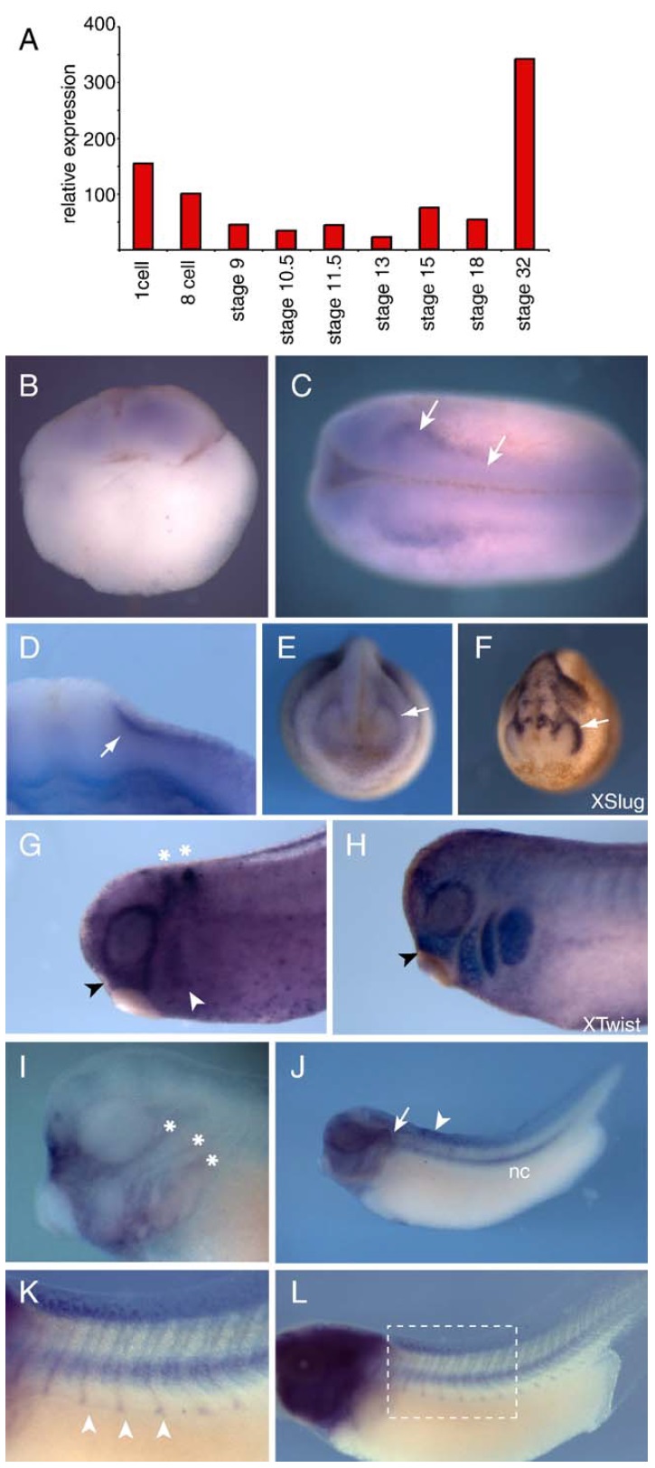
Fig. 2. Expression of Xtes during early Xenopus development. (A) Real-time RTPCR
based expression profile of Xtes from stage 1 through to tail bud stage 32.
Values have been normalized to those of ornithine decarboxylase (ODC). (BâL)
In situ hybridization analysis of Xtes expression. (B) 4-cell stage embryo. (C)
Stage 18; arrows point to strong expression of Xtes next to the neural folds. (D)
Cross-section at neurula stage showing Xtes expression between the epithelium
and mesoderm. (EâH) Xtes expression is found in an anterior stream of cranial
neural crest. (E) White arrow marks the stream of Xtes expressing cells, whose
pattern is similar to the most rostral stream of XSlug positive neural crest cells (F;
white arrow). (G) Xtes expression ventral to the eye (black arrowhead)
corresponds to the most anterior cranial neural crest that is positive for Xtwist
expression (black arrowhead in panel H). White arrowhead and asterisks in
panel G denote Xkrox20 expression in rhombomeres 3 and 5 and the anterior
branchial crest, respectively. (I) Expression of Xtes in the head, in an embryo in
which BM purple stain development was stopped early; asterisks mark the three
lateral line placodes. (J) View of a whole tail bud embryo showing strong
expression in the notochord (nc). Staining was also evident in the otic vesicle
(arrow) and the rostral region of the dorsal fin (arrowhead). (K, L) If the BM
purple stain is allowed to develop longer, Xtes expression becomes evident
along the somitic boundaries (white arrowheads in the higher magnification
view in panel K). Image published in: Dingwell KS and Smith JC (2006) Copyright © 2006. Image reproduced with permission of the Publisher, Elsevier B. V.
Image source: Published Larger Image Printer Friendly View |