XB-IMG-151899
Xenbase Image ID: 151899
|
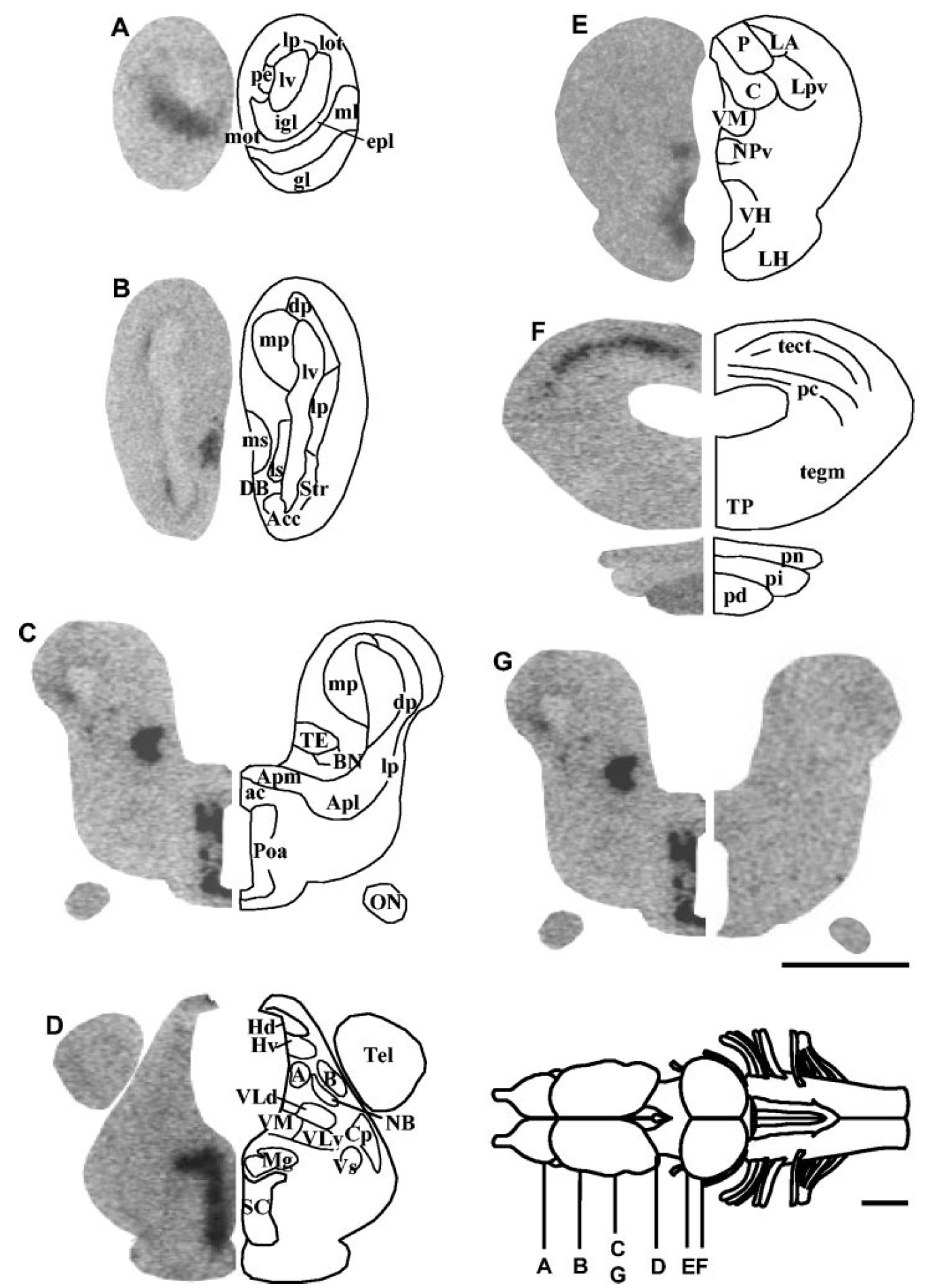
Fig. 1. In situ hybridization histochemistry showing the distribution
of TRH precursor mRNA in the brain and pituitary of Xenopus
laevis. AâF: Frontal brain sections from white-adapted frogs were
hybridized with an antisense TRH precursor riboprobe and exposed
onto Hyperfilm max for 8 days. The anatomical structures, identified
by microscopic analysis, are designated on the right hemisections.
G: A control section incubated with a sense riboprobe (right hemisection)
is compared with a consecutive section hybridized with the
antisense probe (left hemisection). The rostrocaudal levels of the
sections are indicated on the schematic representation of the brain.
Abbreviations as in Table 1. Scale bars 1 mm. Image published in: Bidaud I et al. (2004) Copyright © 2004. Image reproduced with permission of the Publisher.
Image source: Published Larger Image Printer Friendly View |