XB-IMG-152460
Xenbase Image ID: 152460
|
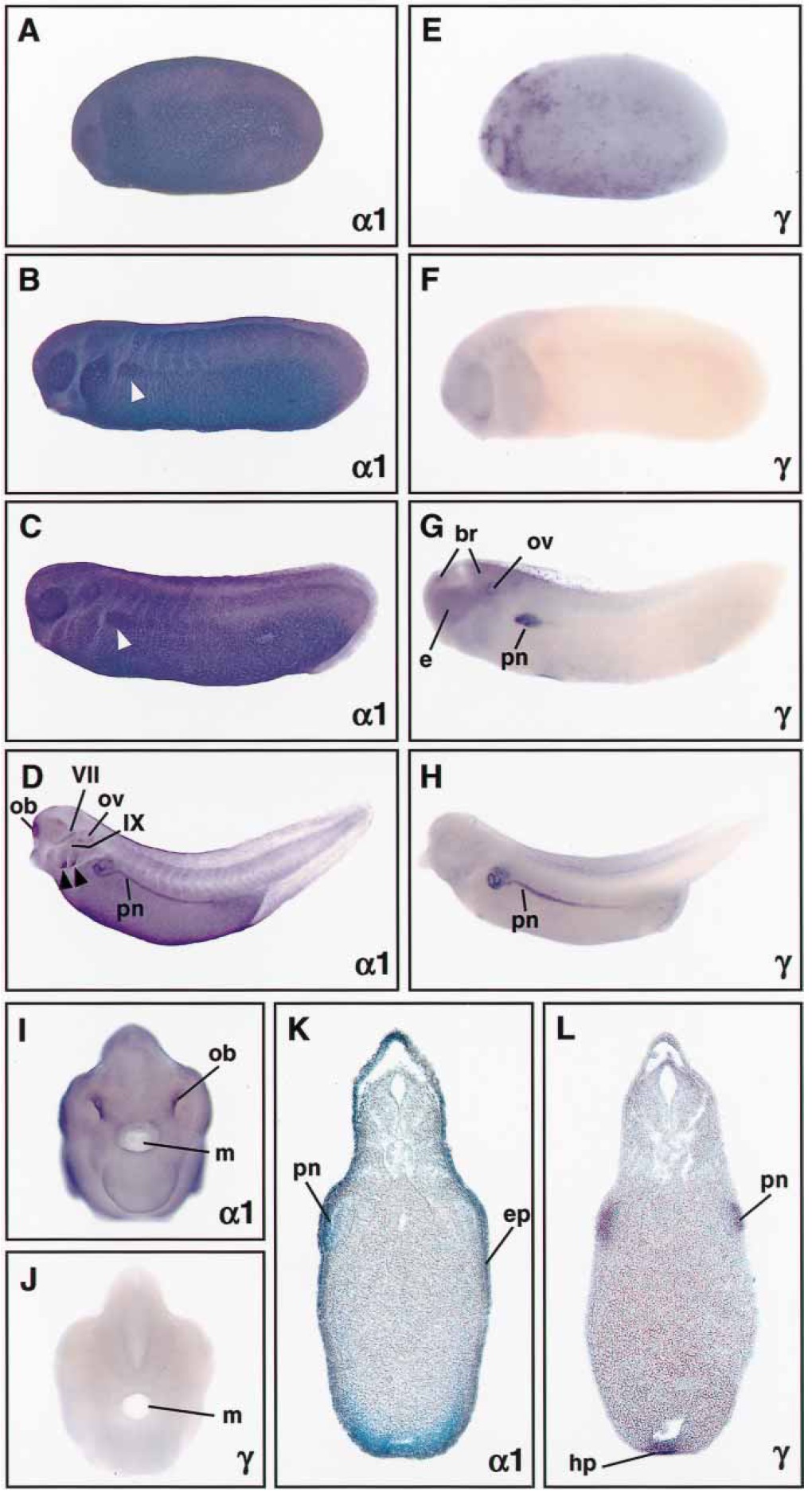
Fig. 3 Expression of Na,KATPase
a1 and g subunit genes
during Xenopus embryogenesis.
Whole-mount in situ hybridizations
were performed
with antisense probes for a1
and g subunits. Transverse sections
of stage 28 (K, L) were
cut at 30â40 mm. Lateral views
(A-H) are shown with anterior
to the left. Frontal views (I, J)
and sections (K, L) are
oriented with dorsal to the top.
A-D a1 subunit expression was
found throughout the epidermis
of tailbud embryos (A,
stage 23; B, stage 26; and C,
stage 28). Expression in the
pronephric anlage can be anticipated
from stage 26 on
(white arrowheads). At stage
37, a1 transcripts were prominently
detected in the olfactory
bulbs, otic vesicles, the gills (arrowheads),
the pronephric kidneys,
and facial (VII) and
glossopharyngeal (IX) nerves.
E-H Stage 22 embryo (E)
showed a punctate staining
pattern for the g subunit. At
stage 25 (F), g expression was
overall low, but became at stage
28 (G) apparent in the brain,
eyes, otic vesicles, and
pronephric primordia. In the
stage 38 embryo (H), strong expression
of g transcripts was
confined to the pronephric
kidneys. I, J Frontal views of
stage 37/38 embryos stained for
a1 and g expression. Strong
staining for a1 transcripts (J)
was seen in the olfactory bulbs.
K, L Transverse sections of
stage 28 embryos revealed a1
(K) in the epidermis and g (L)
expression in the pronephric
and hepatic primordia. For
abbreviations, see legend to
Fig. 2. Image published in: Eid SR and Brändli AW (2001) Copyright © 2001. Image reproduced with permission of the Publisher and the copyright holder. This is an Open Access article distributed under the terms of the Creative Commons Attribution License.
Image source: Published Larger Image Printer Friendly View |