XB-IMG-154152
Xenbase Image ID: 154152
|
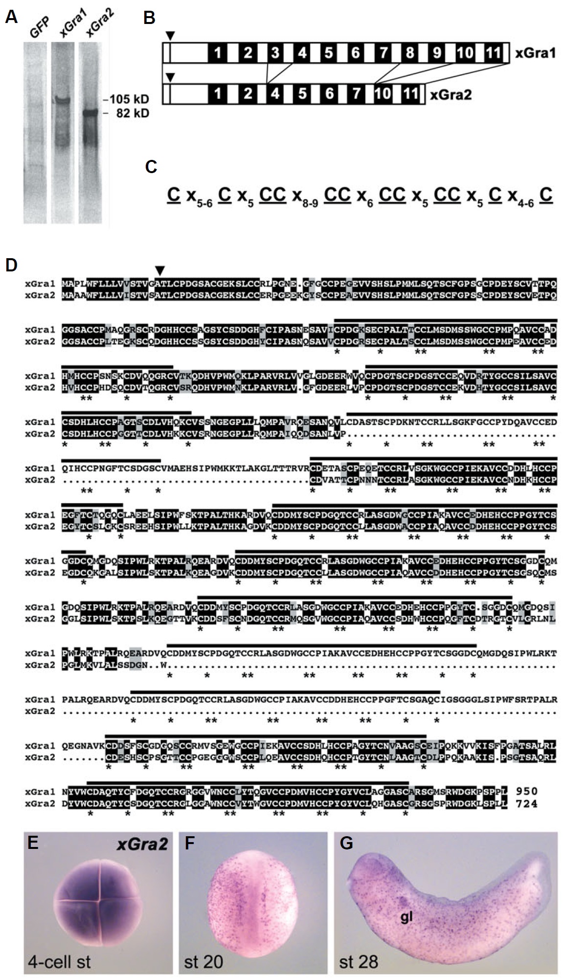
Fig. 3. xGra1 and xGra2 belong to the
Granulin family of secreted growth
factors. (A) Supernatant culture medium
of human embryonic kidney (293T) cells
transfected with cDNA encoding nonsecreted
green fluorescent protein as
control (GFP), Xenopus Granulin-1 (xGra1)
or Xenopus Granulin-2 (xGra2). Cells
were labeled with 35S-methionine and -
cysteine and their supernatants were
analyzed by SDS-PAGE and autoradiography.
Note that xGra1 is secreted as a
105 kD protein and xGra2 as an 82 kD
protein. (B) Diagrams of xGra1 and xGra2.
The signal peptide cleavage sites are
indicated by triangles. The black boxes
indicate conserved granulin repeats (Pfam
accession number PF00396). The numbers
and guidelines indicate repeats conserved
in both proteins and are based on
sequence similarities. Note that the
granulin repeats 3, 8 and 9 of xGra1 are
missing in xGra2. (C) Signature of the
granulin repeat. Note the conserved spacing
of four cysteine doublets flanked by
two cysteine singletons on each side. (D)
Sequence alignment of xGra1 and xGra2.
xGra1 corresponds to the Granulin provisional
protein sequence previously published
for Xenopus laevis (GenBank accession
number AAH48224) and xGra2 is
novel (GenBank accession number
DQ004683). Identical amino acid residues
are shaded in black and similar or
conserved residues in gray. Dots represent
gaps introduced into the amino acid
sequence in order to obtain optimal alignment.
The signal peptide cleavage site as
predicted by SignalP is indicated with an
arrowhead. Black bars indicate the
granulin repeats and the stars the conserved
cysteine residues. The overall
number of amino acids is indicated at the
end of each sequence. (E-G) Expression
of xGra2 analyzed by whole-mount in
situ hybridization. (E) Four-cell stage
embryo in animal view. Note the high
level of maternal transcripts. (F) Embryo
at late neurula stage in dorsal view showing
spotted expression in the epidermis.
(G) Early tail bud stage embryo in lateral
view. Note expression in the pronephric
glomus (gl). Image published in: Pera EM et al. (2005) Copyright © 2005. Image reproduced with permission of the Publisher, University of the Basque Country Press.
Image source: Published Larger Image Printer Friendly View |