XB-IMG-81905
Xenbase Image ID: 81905
|
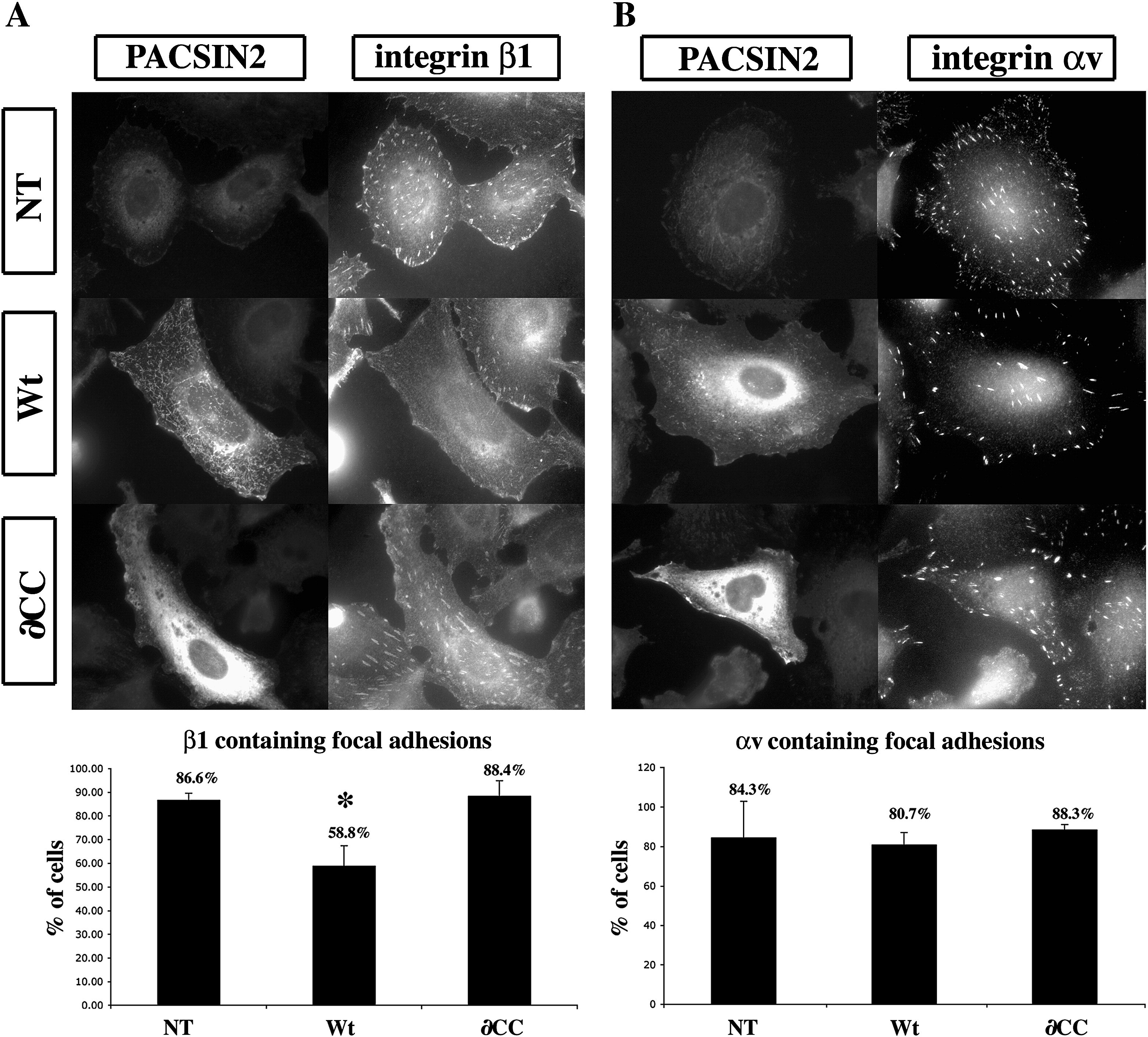
Fig. 8. PACSIN2 over-expression perturbs the recruitment of integrin β1 but not αv to focal adhesion in XTC cells. XTC cells transfected with Wt-PACSIN2 or the âCC mutant were processed for immunofluorescence with a monoclonal antibody against integrin β1 (A) or αv (B) and biotinylated mAb 3D8 against PACSIN2. (A) In non-transfected cells (NT), β1 integrin localizes to focal adhesion (FA). The cells transfected with Wt-PACSIN2 (Wt) display an absence of integrin β1 localization to FA. The expression of the mutant âCC did not affect the localization of β1 integrin to FA (âCC). The percentage of cells displaying integrin β1 positive focal adhesions is plotted on the histogram. The graph represents the mean of three independent experiments. The number of cells scored was as followed: NT = 230, Wt PACSIN2 = 226, âCC = 153. (B) Compared to the non-transfected cells (NT), the localization of integrin αv to FA is unchanged upon expression of Wt-PACSIN2 (Wt) or the âCC (âCC). The histogram represents the percentage of cells displaying integrin αv positive focal adhesions and corresponds to the mean of two independent experiments. The number of cells scored was as followed: NT; n = 148, Wt-PACSIN2; n = 89, âCC; n = 85. The error bar represents standard deviation from the mean. âP < 0.05. Image published in: Cousin H et al. (2008) Copyright © 2008. Image reproduced with permission of the Publisher, Elsevier B. V.
Image source: Published Larger Image Printer Friendly View |