XB-IMG-84186
Xenbase Image ID: 84186
|
||||||||||||||||||||||||||||||||||||||||
|
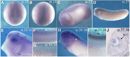
Fig. 1. Spatio-temporal expression pattern of kidins220 mRNA in Xenopus laevis embryos. The developmental stages (st.) are indicated in each panel (A,B) Initial neural tube stage embryos (st. 19), (A) frontal view, dorsal to the top and (B) dorsal view, anterior to the bottom, respectively. (C) Early tail bud stage (st. 22) embryo showing kidins220 expression at the level of the head and in somites. In (A) and (C) the square brackets indicate the forming somites). (D) st. 27 embryo in which the labeled ophtalmic and maxillomandibular branches of the primordium of the trigeminal nerve are highlighted by white and light blue arrowheads, respectively. (E) Magnified view of st.33-34 embryo head, showing signal in forebrain (f), midbrain (m) hindbrain (h), eye vesicle (e), branchial arches (ba). Magnification of the somite region in stage 33-34 embryos, hybridized with Kidins220 (F) and mastr, respectively (G). The square brackets indicate individual somites. (H) Anterior portion of st. 35-36 embryo showing signal in the heart region (red arrowhead) and in otic vesicle (yellow arrowhead). (I) Magnification of somites and dorsal fin of stage 37-38 hybridized embryos. (C-I) Lateral views, anterior to the left, dorsal to the top. (J) Transverse microtome section of hybridized st. 37-38 embryo showing a specific signal in retinal ganglion cells (rgc) and in lens (l). Scale bars: (A-I) 250 um; (J) 50 um. Image published in: Marracci S et al. (2013) Copyright © 2013. Image reproduced with permission of the Publisher, University of the Basque Country Press.
Image source: Published Larger Image Printer Friendly View |